During your visit to Launceston Eye Institute
When you come for your appointment can you please bring:
- Your most recent pair of distance and reading glasses
- A list of your current medications
- Your current Medicare and Pension Health Benefits card
- If you are a contact lens wearer please call in advance to find out if these need to be left out prior to your appointment
- If you wear your contact lenses to the appointment please bring a container to store them in during your examination
- Please ensure your referral is current or has been received at LEI prior to your appointment
- Settlement of your account in full is required on the day of your consultation. Payment by cash, EFTPOS or cheque is available and we can assist you actioning your Medicare rebate on the day.
At most appointments you will have drops that will dilate your pupils, we strongly recommend you arrange to have a driver or alternative transport as your vision will be blurred following your appointment.
At your visit you may be required to have various tests done, some of the tests you may require are explained below.
Angiography (Fluorescein and Indocyanine Green)
An angiogram may be recommended by your Ophthalmologist if any irregularities of the retina are discovered at your consultation. The photographs can then help your specialist make a diagnosis. Ongoing angiograms may also be performed for certain eye diseases to monitor any changes over a period of time.
Age-related Macular Degeneration (AMD) affects millions of people and gradually destroys the sharp, central vision. Central vision is needed for seeing objects clearly and for common daily tasks such as reading and driving. AMD affects the macula, the part of the eye that allows you to see fine detail. An angiograms may be done to show any leaking blood vessels in the macula and can show up any small leaks that could not otherwise be detected.
Angiograms are also beneficial in discovering any leaking blood vessels in the eye associated to Diabetes. Diabetes is a disease in which the body does not properly control the amount of sugar in the blood. Regular eye checks are needed for diabetics, and angiograms can show the Ophthalmologist the exact location of any leaks, they may perform laser treatment to stop the bleeding.
Two different dyes can be injected into the vein: Fluorescein or Indocyanine Green (ICG).
- Fluorescein Angiography:
Most angiograms are done using the Fluorescein dye. Your skin will turn yellow for several hours after the Fluorescein dye is injected. The dye will be flushed out of the system through your urine, which can be a very orange colour for up to 24 hours; drinking water will flush out the dye faster. - Indocyanine Green (ICG) Angiography:
The Indocyanine Green (ICG) angiogram is a similar but less frequently performed investigation. An ICG angiogram can sometimes locate abnormal choroidal vessels better than a fluorescein angiogram however an ICG is not recommended for patients with an allergy to iodine.
What should I expect?
Digital cameras are used to take photographs of the eye. The advantages of using a digital camera are that only half the amount of dye is required for the angiogram reducing the adverse affects associated with the dye.
Sometimes the cannula used to inject the dye into the arm can damage a fragile vein and can cause burning and yellowing of the skin around the injection point. This usually only lasts a very short time and the yellowing on the skin will go away after a few days.
When the dye is injected into the vein, some patients experience nausea, this passes after a few seconds. Allergic reactions to the dye are rare. Rashes or skin irritations are treated with oral or injected antihistamines. Reactions of blood pressure falling are extremely rare.
Fluorescein is reported to be safe in pregnancy, but we prefer to avoid this test in pregnant patients where possible. Please advise the staff prior to the test being commenced if you are or suspect you may be pregnant. If you have any questions please ask prior to the test.
Biometry
You may have a biometry screening done if you have cataracts. The biometry measures the length and curvature of your eye. At the time of surgery, the lens in your eye is removed and replaced with an artificial lens. The biometry test tells us the strength of the lens required for you. If you have a biometry you will also have Topography & Autorefraction. A Medicare refund is available for this testing procedure.
Hard contact lens wearers should leave their contact lens out for at least two weeks prior to their consultation and soft contact lens wearer for at least one week.
Computer Perimetry (Field)
A computer perimetry (also known as a visual field) tests the entire scope of your peripheral (side) vision.
This test is frequently used to detect any sign of glaucoma damage to the optic nerve. In addition, perimetry tests are also useful for detecting central or peripheral retinal disease, eyelid conditions such as ptosis or drooping, optic nerve disease and disease affecting the visual pathways within the brain.
The perimetry test is performed one eye at a time with the other eye completely covered to avoid errors. You will be asked to look directly ahead at all times to avoid testing the central vision rather than the peripheral vision.
On your first visit to the ophthalmologist, with signs of glaucoma, the ophthalmic technicians will perform a perimetry test collating the results for the ophthalmologist to view during your consultation. This initial test is commonly referred to as your ‘base line’ test. This enables the ophthalmologist to form a starting point and then at regular intervals, further perimetry tests are performed and comparisons are made against your initial test. Without regular 6 monthly tests it is very difficult for the ophthalmologist to accurately monitor your progress. Multiple 6 monthly tests are a very important part of assessing and monitoring your condition as it can detect any subtle changes in your vision. Some medications you take can also affect your central vision (such as Plaquenil) so you may need to have more regular field tests for this reason.
In some patients, not all, this 6 monthly testing may be co-managed between the ophthalmologist and your optometrist.
Currently a Medicare refund is available for this test.
Disc or Retinal Photos
Optic disc photography is important as it helps create a baseline for future comparison. Your ophthalmologist may later take additional pictures to see if there are any changes to the nerve fibre layer and associated thinning of the tissue at the optic nerve head. These images can help identify signs of glaucoma progression.
You may have retinal photos taken if you have changes on the retina such as choroidal nevus (freckle). This allows your doctor to compare the photos over a long period of time to monitor any change. Medicare currently do not offer a refund on this testing procedure.
Optical Coherence Tomography (OCT)
Optical Coherence Tomography is a non-invasive, non-contact imaging technique used to take images of the retina and the optic nerve. By focusing beams of light into the eye it will scan a cross section image, similar to a topographical map.
This machine assists greatly in diagnosing and monitoring macular diseases including Age-related macular degeneration, macular holes, macular oedema, epiretinal inflammatory diseases and it also assists in the assessment of glaucoma as it is able to assess the nerve damage to the back of the eye.
There is a fee charged for the OCT test which currently Medicare do not offer any rebate on. Depending on the patient’s level of cover, some private health funds will rebate a portion of the fee under ancillary cover.
Pachymetry
Pachymetry is a simple, quick and painless test to measure the thickness of the cornea and is essential for patients with corneal abnormalities as well as patients with glaucoma. When combined with standard intraocular pressure measurements, pachymetry gives your ophthalmologist a much more accurate prediction of glaucoma development. Currently Medicare does not offer a refund for this test.
Scheimpflug
The Scheimpflug is a rotating camera which captures images of the anterior eye segment. A Scheimpflug measurement process takes less than two seconds to measure each eye, and then generates images and corneal maps used for diagnosis and treatment of cataracts, glaucoma and corneal problems. Currently Medicare do not offer any refund for this test.
Specular Microscopy
Specular microscopy is a non-invasive photographic technique that allows you to visualise and analyse the corneal endothelium. This test is used to monitor the number, density, and quality of endothelial cells that line the back of the cornea. A microscope magnifies the cells thousands of times and the image is captured with a camera or video camera. The number of cells within one square millimeter are counted and recorded. The endothelium of a young, ten-year-old with a healthy cornea has approximately 3,500 cells in each square millimeter. Normal aging causes the cells to gradually decrease over time. By age 60, most people have approximately 2,500 cells per square millimeter.
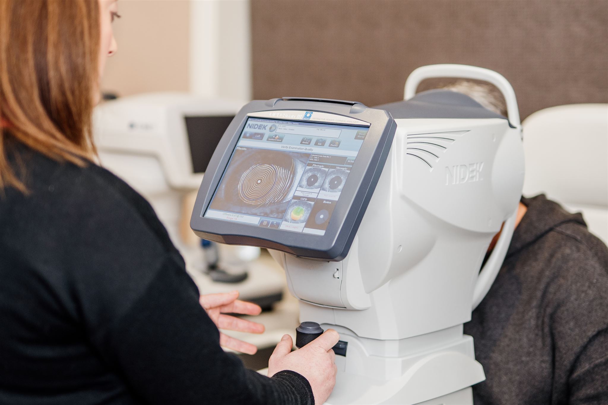
Topography & Autorefraction
Topography & Autorefraction are done at the same time. Topography is a topographical map of your cornea. You may have this done if you have corneal trouble such as Keratoconus or if you have cataracts. Autorefraction provides an objective measurement of your refractive error (the strength glasses you wear). It is particularly important to know your refraction prior cataract surgery to calculate the most suitable artificial lens. Currently Medicare does not offer a refund for this vital test.

